
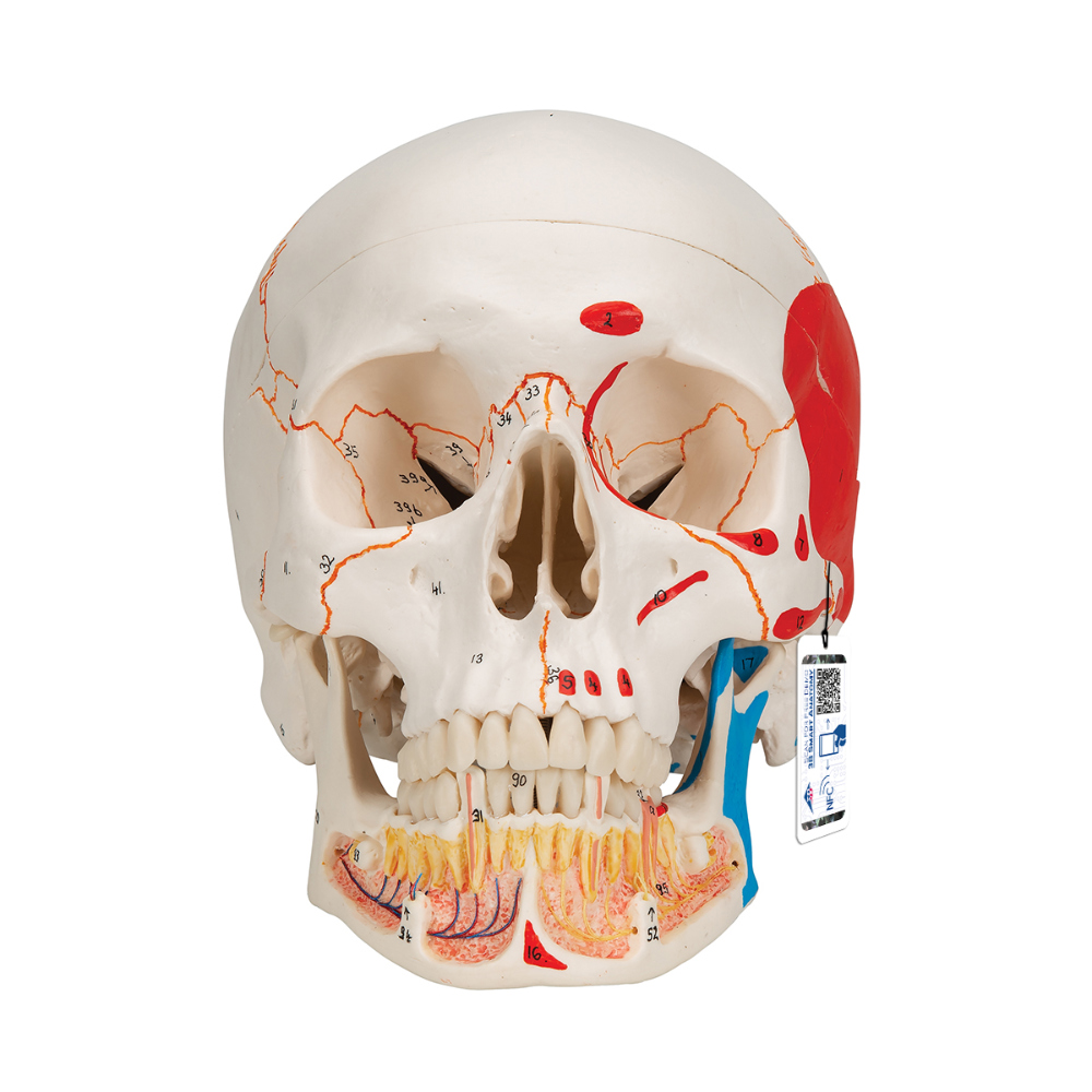
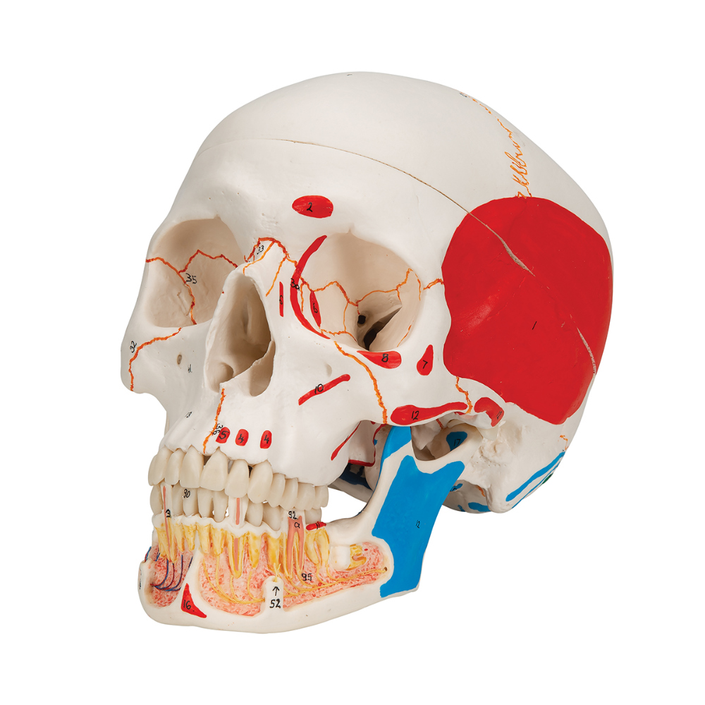
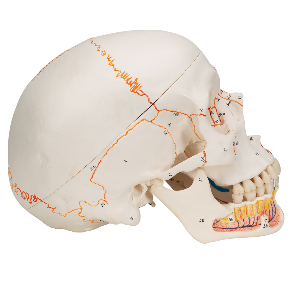
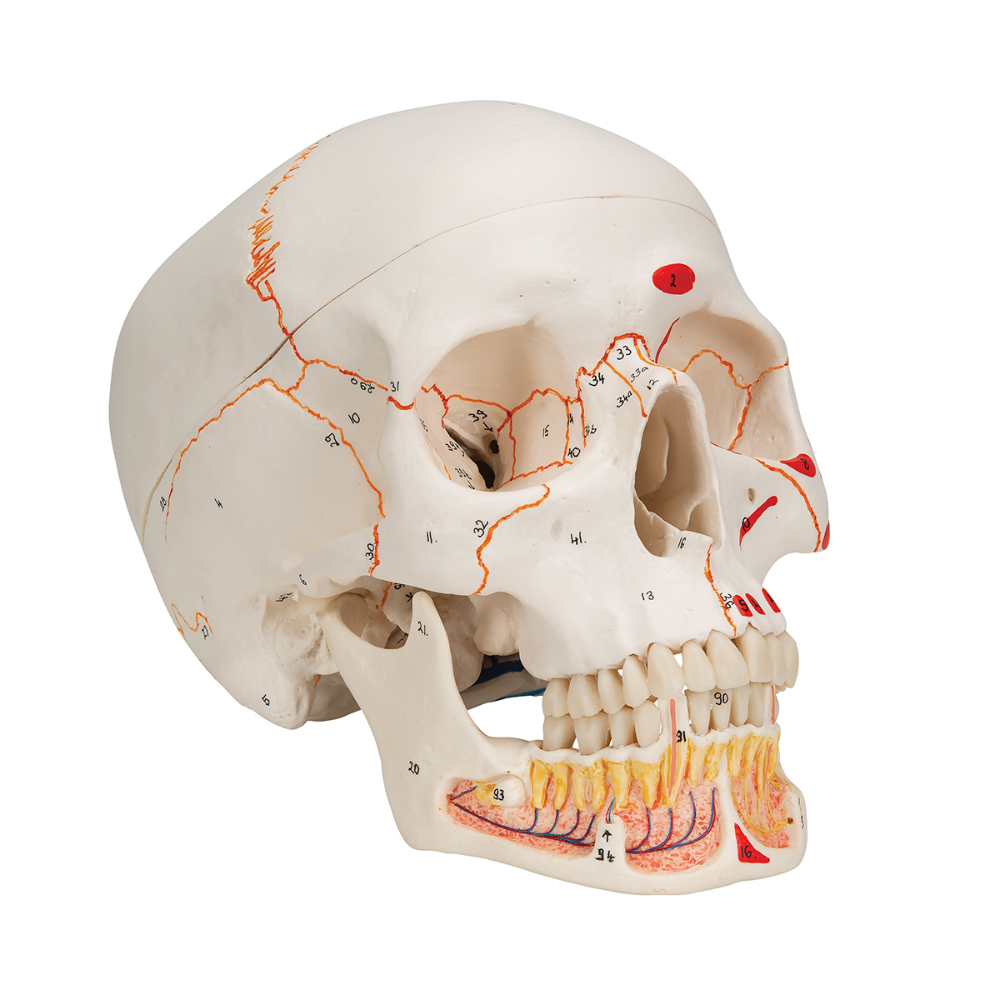
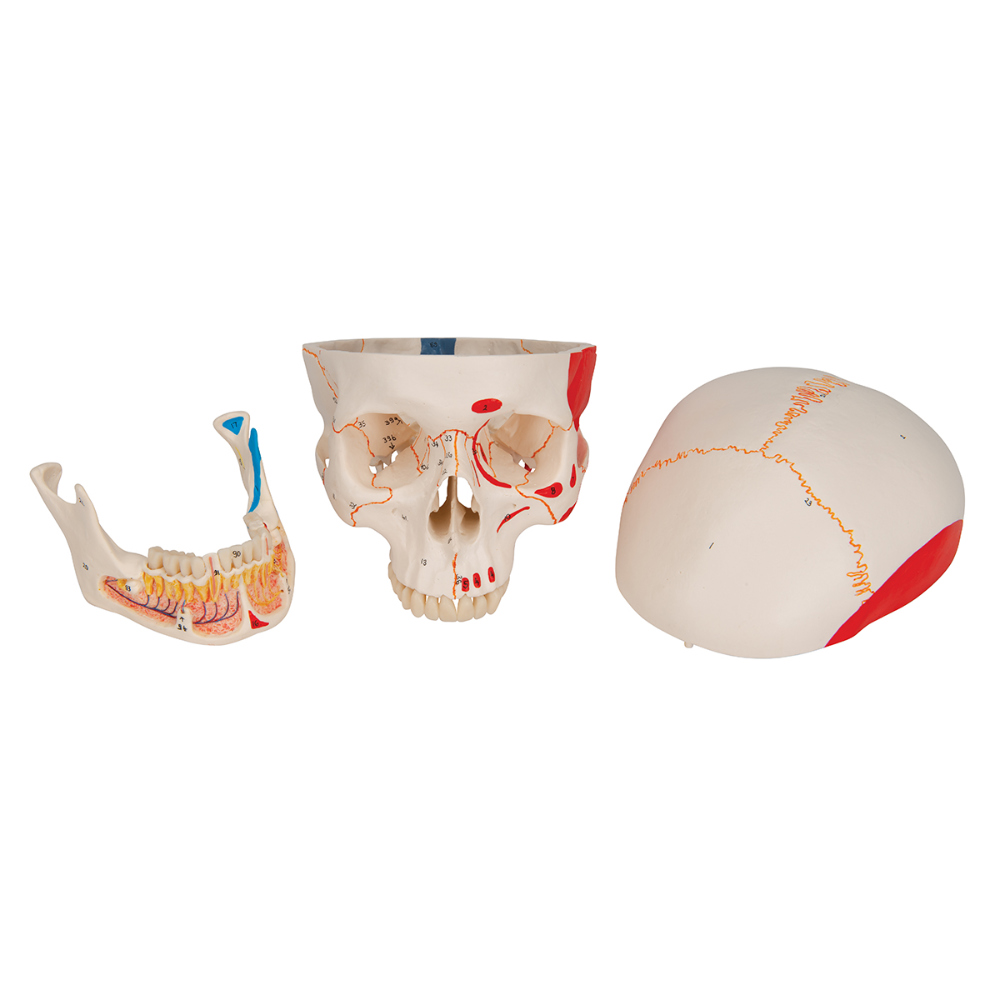
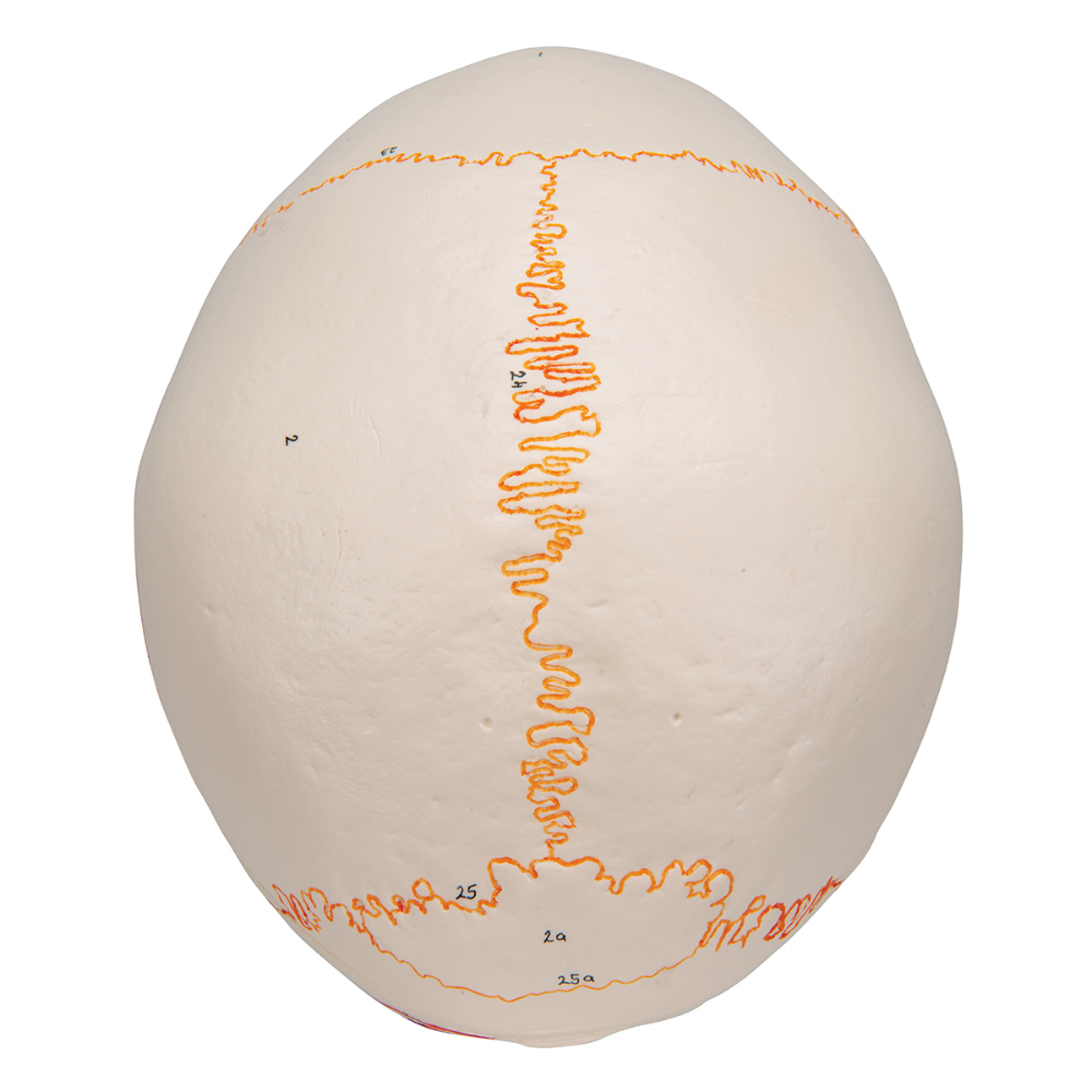
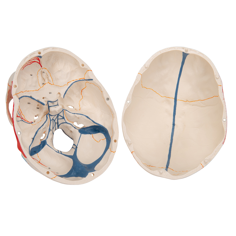
| 상품명 | [특별할인] 하악 노출 채색된 두개골모형 3파트 분리형 Classic Human Skull Model with Opened Lower Jaw A22/1 [1020167] |
|---|---|
| 판매가 | 별도문의 |
| 국내·해외배송 | 국내배송 |
| 배송방법 | 택배 |
| 배송비 | 4,400원 (100,000원 이상 구매 시 무료) |
| 원산지 | EU |
| SNS 상품홍보 | |
|---|

| 옵션선택 |
(최소주문수량 1개 이상 / 최대주문수량 0개 이하)
사이즈 가이드수량을 선택해주세요.
위 옵션선택 박스를 선택하시면 아래에 상품이 추가됩니다.
| 상품명 | 상품수 | 가격 |
|---|---|---|
| [특별할인] 하악 노출 채색된 두개골모형 3파트 분리형 Classic Human Skull Model with Opened Lower Jaw A22/1 [1020167] |


|
별도문의 ( |
할인가가 적용된 최종 결제예정금액은 주문 시 확인할 수 있습니다.
![[특별할인] 하악 노출 채색된 두개골모형 3파트 분리형 Classic Human Skull Model with Opened Lower Jaw A22/1 [1020167]](http://3bs.kr/web/product/big/202201/b571f0743213fb0d8e6af8198124df57.jpg)


게시물이 없습니다
게시물이 없습니다

