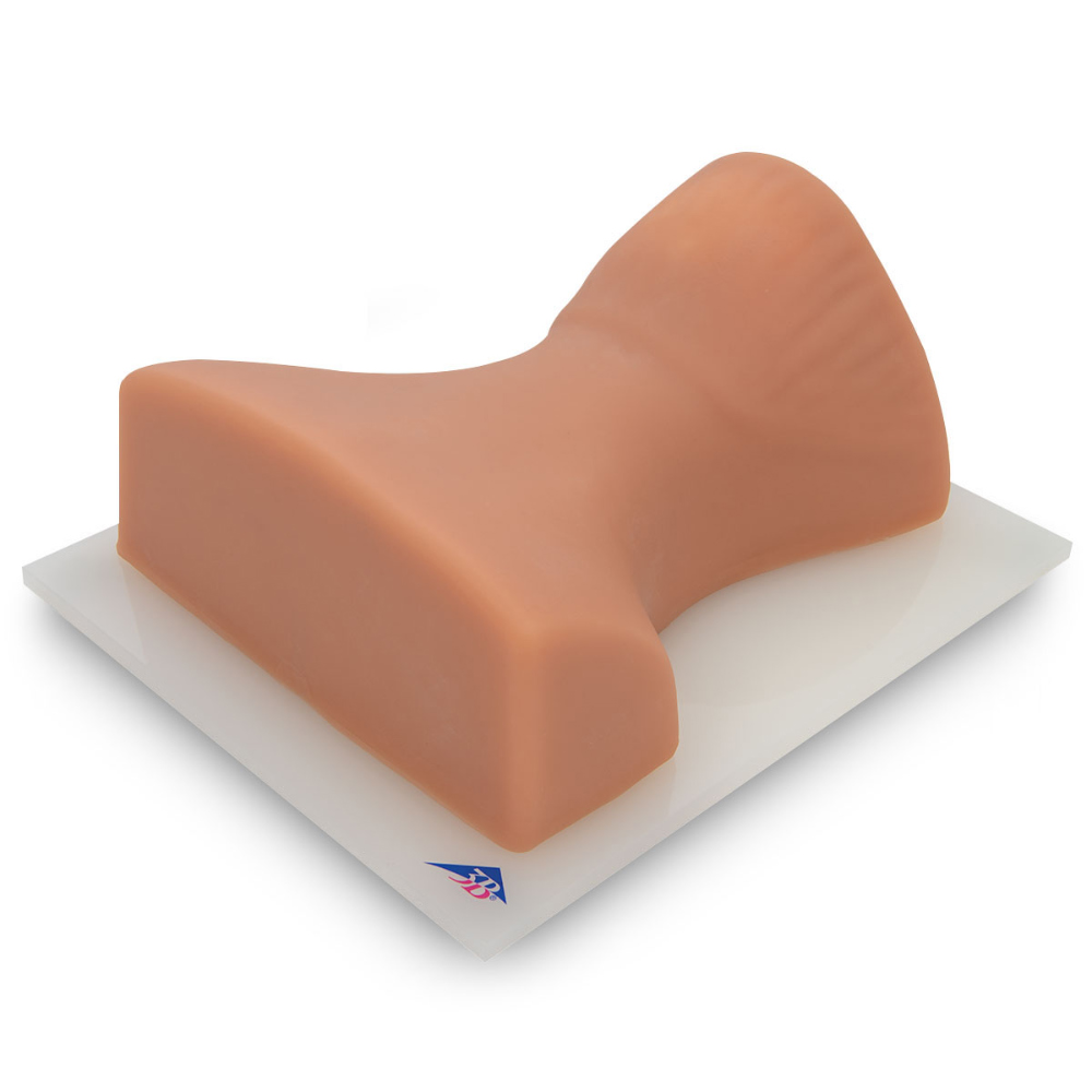
이미지 유도 경추 주사 트레이너
Image Guided Cervical Spine Injection Trainer P67
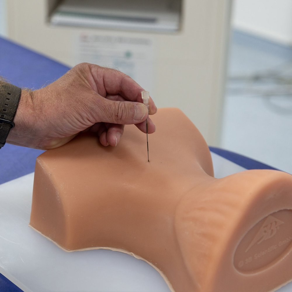
실제와 같은 방사선량을 가진 경추에 대한 사실적인 영상 유도 주사 훈련.
영상 유도 경추 주사 트레이너는 연습생이 성공적인 척추 주사 절차에 대한 차원적 이해의 3가지 방법을 발전시킬 수 있도록 합니다.
그들은 중재적 통증 절차와 관련된 경추 해부학과 영상과의 상관관계를 배우고, 영상 및 해부학적 검사를 사용하여 대상 조직와 취약한 구조를 식별하며, 중재적 척추 시술 중 환자의 안전을 보장하기 위한 모범 사례를 적용하는 방법을 배울 수 있습니다.
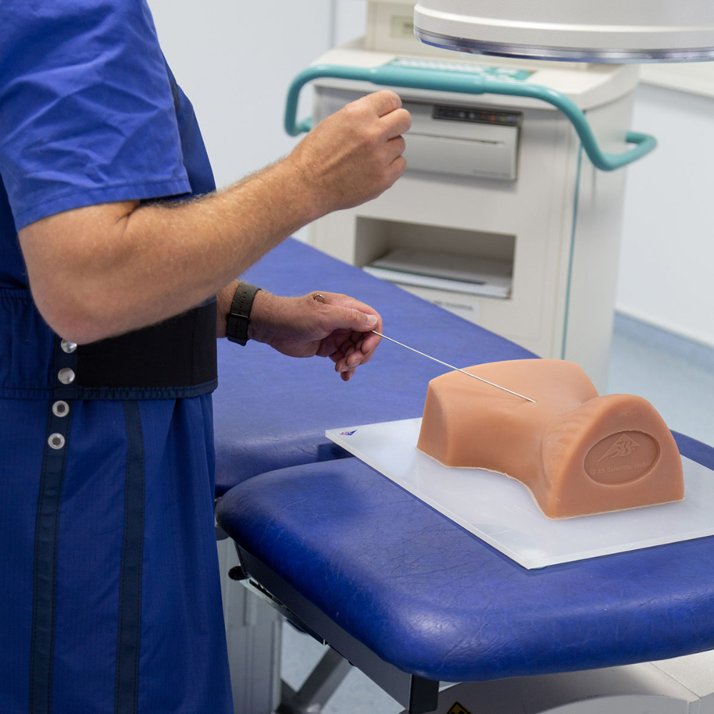
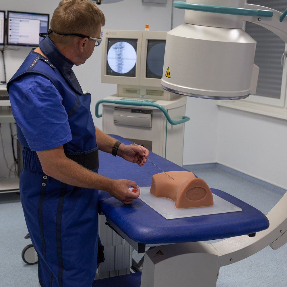
척추 주사 트레이너를 사용하는 것은 영상 강좌와 생체 기술 연구소의 경제적인 대안입니다.
신뢰할 수 있고 표준화된 환자 시뮬레이션이 항상 준비된 상태로 강사에게 제공합니다.
• 사실적은 X선 영상을 위한 실제와 같은 방사선량
• 사실적인 주사 요법
• 해부학적으로 정확한 골격 구조
• 육안으로 식별 및 촉진 가능한 랜드마크
시뮬레이터에서 다음과 같은 영상 유도 척추 주사 교육을 할 수 있습니다.
• 랜드마크 유도 개입 기법
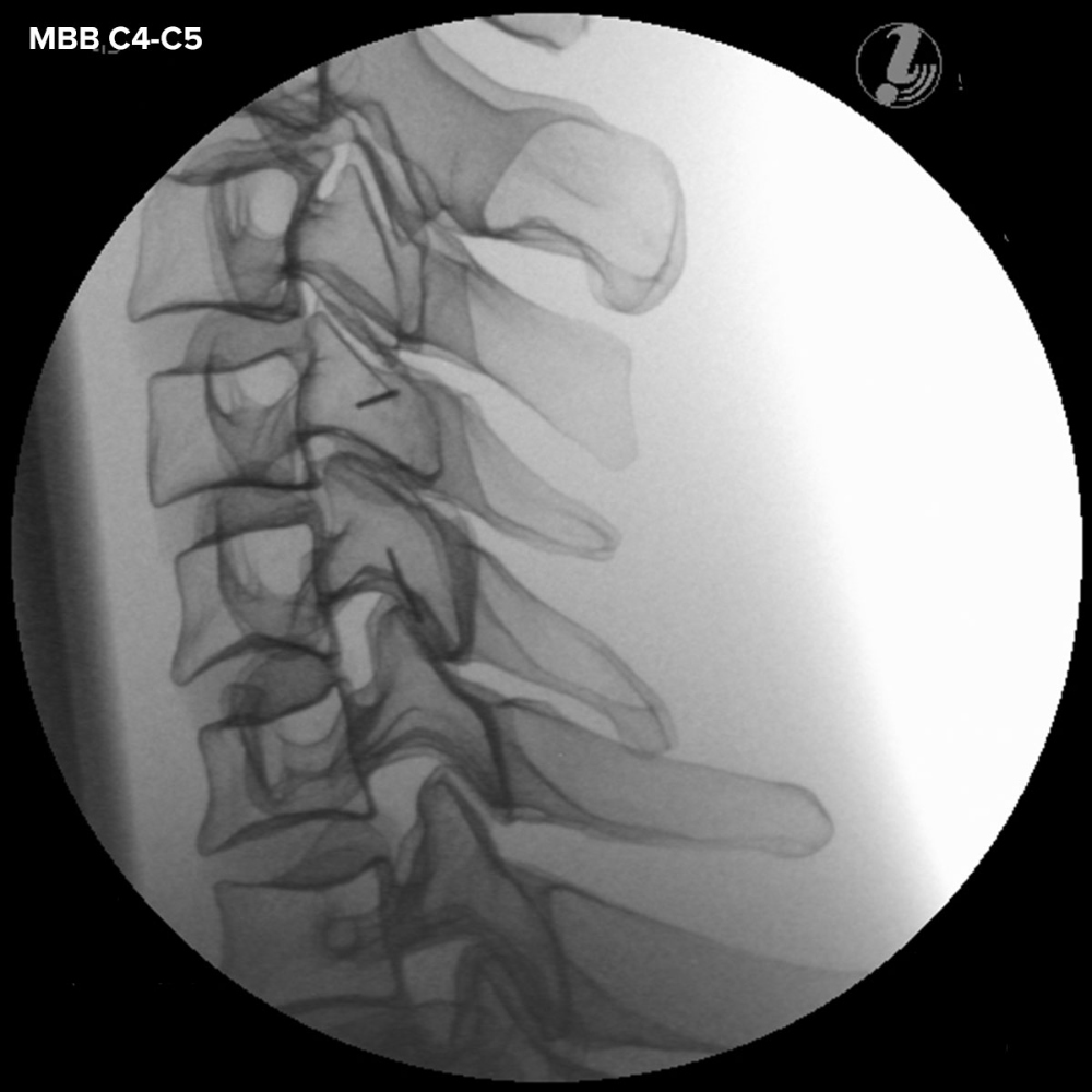
• 중간 분기 블록
• 후두부 블록
• 측면 고주파 제신경
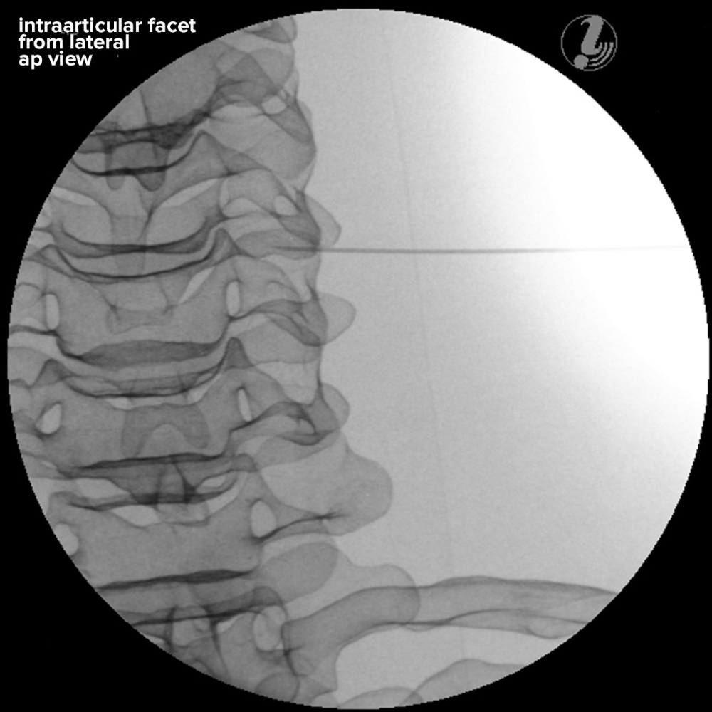
• 관절 내 C1/C2
• 관절 내 경추 후지
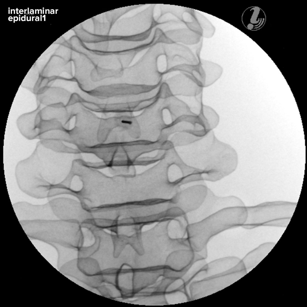
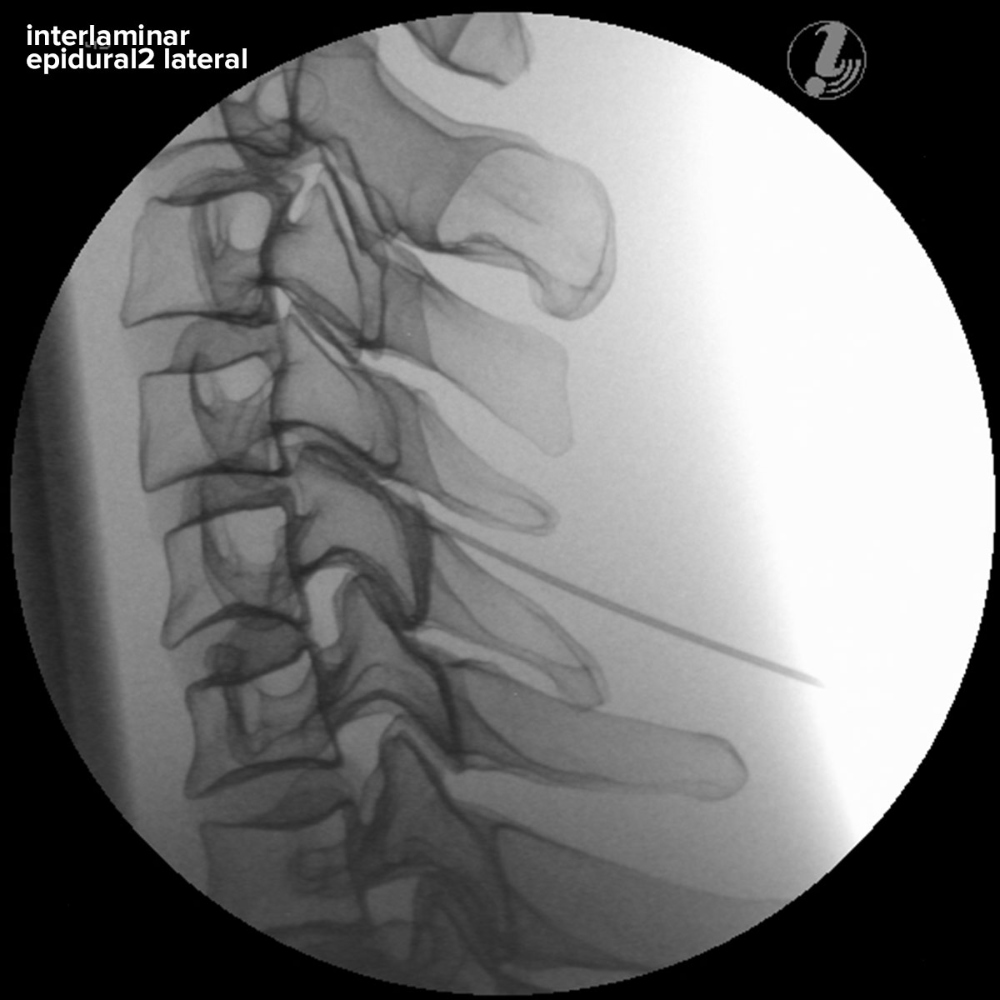
• 추궁판간 경막 외 주사
경추 주사 시뮬레이터는 다움과 같은 뼈 구조를 보여줍니다.
• 후두골(외후두 돌기 두개)
• 척추골 C1-Th2
• 늑골 1과2
트레이너는 다음과 같은 기술적 특성 덕분에 이미지 코스에서 특히 적합합니다.
• 자동 봉합 소재는 주사 실습에 반복적으로 사용할 수 있습니다.
• 운반함으로 휴대가 용이함
• 내구성이 좋고, 청소가 쉬움
구성품
• 경추 주사 트레이너 P67 이미지 안내서 1개
• 안전한 운반함 1개
• 보호 커버 1개
• 탤컴파우더 용기 1개
• 2.73 kg
Realistic image guided injection training on the cervical spine with life-like radiopacity
The Image Guided Cervical Spinal Injection Trainer enables trainees to develop a three-dimensional understanding of the procedures for successful spine interventions. They will learn to correlate imaging with the cervical spinal anatomy relevant to interventional pain procedures, to identify the target tissue and vulnerable structures using imaging and anatomic inspection, and to apply best practices to ensure patient safety during the interventional spine procedures.
Using Spinal Injection Trainers is an economic alternative for imaging courses and bio-skills labs on cadavers and offers the instructors a reliable, standardized patient simulation always ready to use:
• Life-like radiopacity for realistic x-Ray images
• Realistic injection haptics
• Anatomically accurate bone structure
• Visually identifiable and palpable landmarks
The following image guided spinal interventions can be trained on the simulator:
• Landmark Guided Intervention Techniques
• Medial Branch Block
• Occipital Block
• Facet Radiofrequency Denervation
• C1/C2 Intraarticular
• Cervical Facet Intraarticular
• Interlaminar Epidural Injection
The Cervical Spinal Injection Trainer depicts the following bone structures:
• Occipital bone (cranial to external occipital protuberance)
• Vertebrae C1-Th2
• Ribs 1 and 2
The trainer is especially suitable for use in imaging courses thanks to these technical features:
• Self-Sealing material can be used repeatedly for injection training
• Completely portable with a secure transport box
• Durable construction, easy to clean
Delivery content:
• 1 Image Guided Cervical Spinal Injection Trainer P67
• 1 secure transport box
• 1 protective cover
• 1 container of talcum powder


![이미지 유도 경추 주사 트레이너 Image Guided Cervical Spine Injection Trainer P67 [1021900]](http://3bs.kr/web/product/big/202204/d2a1ec419929f349d9f099448f4b39e0.jpg)